Most of my MSc thesis used a lot of math and fancy-dancy computer modeling to look at whether it is biologically feasible for ankylosaurids to have used their tail clubs for forceful impacts (and therefore as offensive or defensive weapons). But another way to look for evidence of behaviour is to look for injuries, which can sometimes, if you’re lucky, give you clues about some of the more dramatic moments in an animal’s life. So as I was looking at specimens for my MSc (and into my PhD), I always kept an eye open for anything unusual or abnormal that could be a pathology.
What I would love to tell you is that I found healing fractures in the handle vertebrae or knob of the tail club, because that would be pretty good evidence that ankylosaurids were smashing things with their tails. Sadly, I have not seen anything like that in any of the tail club specimens I’ve seen. There are several explanations for the absence of handle pathologies:
- Ankylosaurids did not use their tails for forceful impacts.
- Ankylosaurids did use their tails for forceful impacts, but the impact forces were never enough to cause the tail to break.
- Ankylosaurids did use their tails for forceful impacts, and the impact forces were so great that a fracture was probably catastrophic and unlikely to heal.
- Ankylosaurids did use their tails for forceful impacts, and injuries occurred sometimes, but we haven’t found any injured specimens yet.
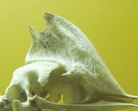
What else did we find? Well, there’s a tail club knob that may have become infected – it’s on display at the Royal Tyrrell Museum Field Station if you happen to be visiting Dinosaur Provincial Park this summer, and you can check it out for yourself. There's a big hole on the right major osteoderm, and this osteoderm is also much taller in posterior view.
And finally, several specimens of anterior free caudal vertebrae had rough, rugose bone textures on the neural spines and sometimes on the transverse processes. In two specimens (a Euoplocephalus at the AMNH and the Talarurus mount at the Palaeontological Institute in Moscow) some of these anterior vertebrae are fused together. At first I thought these might be infections, but as it turns out, the anterior caudal vertebrae of whales and crocodiles can fuse as a mechanical response to stress from swimming motions (see Galatius et al. 2009 and Mulder 2001). It is conceivable that this was occurring in ankylosaurids either as a response to keeping the heavy tail club elevated off that ground, or as a result of tail club swinging.
I wasn’t able to draw any definitive conclusions about tail-clubbing in ankylosaurs based on my pathology survey, and trying to diagnose bone pathologies in fossils is EXTREMELY challenging. Perhaps it is something that gets easier with time. But I had a lot of fun with this project and I think it is worthwhile to look at palaeopathologies as potential sources of information, as long as you’re careful how you interpret them.
References!
Arbour VM, Currie PJ. 2011. Tail and pelvis pathologies of ankylosaurian dinosaurs. Historical Biology, online 19 May 2011.
Galatius A, Sonne C, Kinze CC, Dietz R, Beck Jensen J-E. 2009. Occurrence of vertebral osteophytosis in a museum sample of whitebeaked dolphins (Lagenorhynchus albirostris) from Danish waters. Journal of Wildlife Disease 45:19–28.
Mulder EWA. 2001. Co-ossified vertebrae of mosasaurs and cetaceans: implications for the mode of locomotion of extinct marine reptiles. Paleobiology 27:724–734.
Tanke DH, Farke AA. 2007. Bone resorption, bone lesions, and extracranial fenestrae in ceratopsid dinosaurs: a preliminary assessment. In: Carpenter K (ed). Horns and beaks. Bloomington (IN): Indiana University Press. p. 319–348.
Tanke DH, Farke AA. 2007. Bone resorption, bone lesions, and extracranial fenestrae in ceratopsid dinosaurs: a preliminary assessment. In: Carpenter K (ed). Horns and beaks. Bloomington (IN): Indiana University Press. p. 319–348.


